
Innovation and research for fluorescence microscopy

NOS1000
Optical-Sectioning Microscopy
Introducing the NOS1000, Nexcope's structured-light optical sectioning microscope. Designed for advanced fluorescence imaging where background rejection, resolution, and versatility are critical.
Employing specialised illumination patterns to enhance optical resolution, the NOS1000 surpasses the diffraction limit of conventional fluorescence microscopes. Its variable structured light system enables rapid, gentle optical sectioning for 3D imaging, effectively reducing out-of-focus fluorescence interference and autofluorescence from specimens and culture media. The result is sharper, more authentic sample images - opening up more diverse possibilities for your research.

Structured Light + Computational Imaging
Versatile resolution enhancement
The NOS1000 combines spatial light modulation with computational imaging algorithms to achieve lateral resolution of 120nm (at 405nm) and optical section thickness as fine as 250nm (at 488nm). This places the NOS1000 ahead of comparable systems on both resolution and sectioning performance, at a significantly lower price point.


Perfect integration with NIB1000
Structured Light + Computational Imaging
The NOS1000 combines spatial light modulation with computational imaging algorithms to achieve lateral resolution of 120nm (at 405nm) and optical section thickness as fine as 250nm (at 488nm). This places the NOS1000 ahead of comparable systems on both resolution and sectioning performance, at a significantly lower price point.
Key Features
Built For Discovery

High-Resolution Imaging
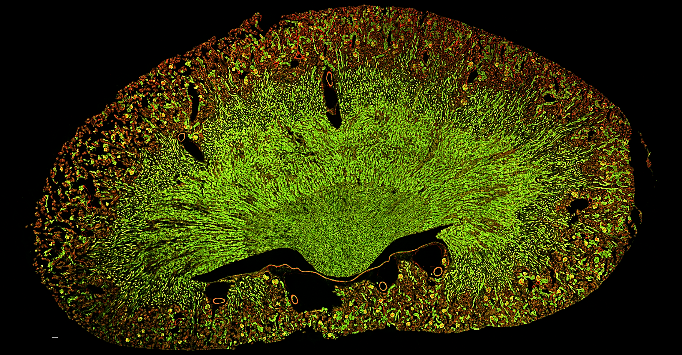
Achieves optical resolution of 120 nm in the XY-plane and 200 nm along the Z-axis, effectively eliminating out-of-focus stray light and clearly revealing fine microstructural details. Slide provided by Argolight.

Optical Sectioning for 3D Imaging
Enables rapid, gentle optical sectioning for 3D imaging, effectively reducing out-of-focus fluorescence interference as well as autofluorescence from specimens and culture media.

Full motorisation and control
High-precision motorised XY stage for automated multi-position and time-lapse experiments. Integrated navigation preview to locate and return to regions of interest.
Variable Structured Light System
Patterns are projected sharply onto the focal plane, while being suppressed on out-of-focus planes. Variable patterns offer greater flexibility and adaptability than conventional fixed structured light systems.

Fast Imaging
High-speed imaging rate of up to 30 FPS. Tracks fast-changing intracellular processes in real time - including protein transport, ion flux, and more.
Multidimensional Imaging
Fully-featured NOMIS Pro X software supports X/Y/Z/λ/T/multi-well acquisition, freely combined to meet diverse and complex experimental applications.


Advanced Optics
Novel Optics designed and manufactured components
Novel Optics, the parent company of Nexcope is one of the most advanced optics companies in the world. Headquartered in Ningbo, China, Novel Optics designs, produces and innovates with the best in optical precision. Enjoy a system featuring the finest optical and mechanical components!
Key Specifications
| Specification | Detail |
|---|---|
| Optics & Observation | |
| Eyepiece | 10×, diopter adjustment -5 ~ +5 |
| Observation Tube | Hinged binocular tube, 10-40° tilt, eye relief 47-78mm, eyepiece interface Φ30 |
| Tube Mount (standard) | Binocular/trinocular switching: (100/0; 0/100) |
| Tube Mount (optional) | 1× C-mount |
| Nosepiece (standard) | Motorised 6-position nosepiece (expansion slot) |
| Nosepiece (optional) | Motorised 6-position nosepiece with anti-focus drift module |
| Bertrand Lens | Switchable into/out of optical path, focus adjustable |
| Intermediate Magnification | 1×, 1.5× switching |
| Stage & Focus | |
| Stage | Motorised; travel range 130mm × 100mm (table size 445mm × 300mm); universal holder for 35-65mm culture dishes and slides; plate holder bracket |
| Focus System | Electric drive, travel 10mm, minimum step 0.02μm, repeatability 0.1μm; 3-speed focus handwheel |
| Display & Control | |
| Touch LCD Display | 5.7-inch built-in touchscreen (front-mounted); displays light source intensity, objective magnification, fluorescence band, intermediate magnification, turret position, Bertrand lens status |
| Main Body Port | 4-speed electric switching; beam splitter - Binocular Right/Left = 100%:0; Left: Binocular = 100%:0; Right: Binocular = 20%:80% |
| Main Body Side Port | 1× C-mount |
| Joystick | XYZ electric control; displays objective magnification and fluorescence band status; shortcut function settings |
| Control Software | NOMIS Pro X Microscope Image Processing Software |
| Illumination | |
| Transmitted (standard) | Köhler LED illumination; field/aperture diaphragms |
| Epi-illumination (optional) | LED light source (385nm, 470nm, 555nm, 630nm); field/aperture diaphragms; 2-hole filter slider; lamp controller; electric shutter (software-controllable) |
| Structured Illumination | Adjustable grid; built-in high-power long-life LED projection module (405nm, 470nm, 560nm, 630nm) |
| Condenser | 7-position electric turret; 4 small 37mm openings (phase contrast, Hoffman modulation, ND filters); 3 large 39mm openings (DIC, ND filters); travel range 66mm; NA=0.52; WD=30mm |
| Fluorescence | |
| Fluorescence Turret | 6-position motorised turret (FITC, TRITC, DAPI standard); Φ25/30 light transmission; electric shutter |
| Fluorescence Second Layer (optional) | Z-axis elevated base, fluorescence turret elevated base, stage elevated base |
| DIC Accessories (optional) | All magnifications |
| Camera | |
| Camera (standard) | sCMOS back-illuminated monochrome; 2048×2048 pixels (4MP); pixel size 6.5μm×6.5μm; chip 13.3mm×13.3mm; QE 95%@600nm; 100fps@USB 3.0 |
| Camera (optional) | sCMOS back-illuminated monochrome; ≥3200×3200 pixels; pixel size 6.5μm×6.5μm; chip 20.8mm×20.8mm; peak QE >95%; 83fps@full frame (16bit) |
Downloads