
Innovation and research for fluorescence microscopy

NCF2000
Confocal Microscope
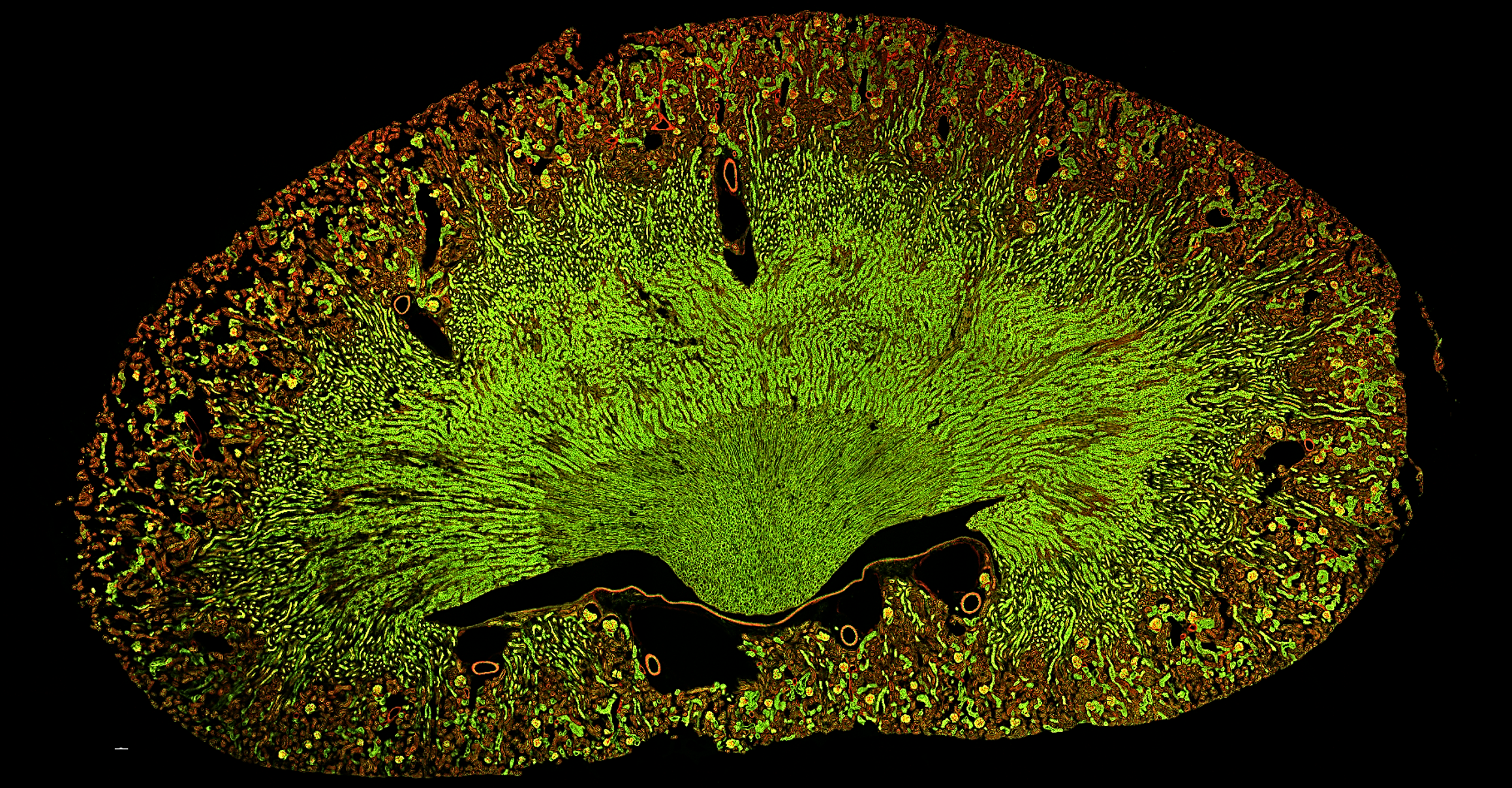
Introducing the NCF2000, Nexcope's next-generation laser confocal microscope, designed for high-throughput and high-quality imaging in modern biological research.
Featuring an industry-leading 25mm field of view, resonant scanning technology, and a high-sensitivity GaAsP detection system, the NCF2000 is purpose-built for large samples, live cells, and demanding multi-channel experiments.
Advanced technology for cutting-edge research
The NCF2000 pairs with the NIB1000-AT inverted microscope to form a comprehensive confocal imaging platform. With scanning resolution up to 8192 x 8192 pixels and real-time four-channel synchronised imaging, the system handles everything from large-area tissue mapping to rapidly changing live-cell processes.
Expandable functions include a live cell culturing system, super-resolution structured illumination microscopy (SR-SIM) module, and dual-layer fluorescence accessory - adapting to the evolving demands of cutting-edge research across disciplines.

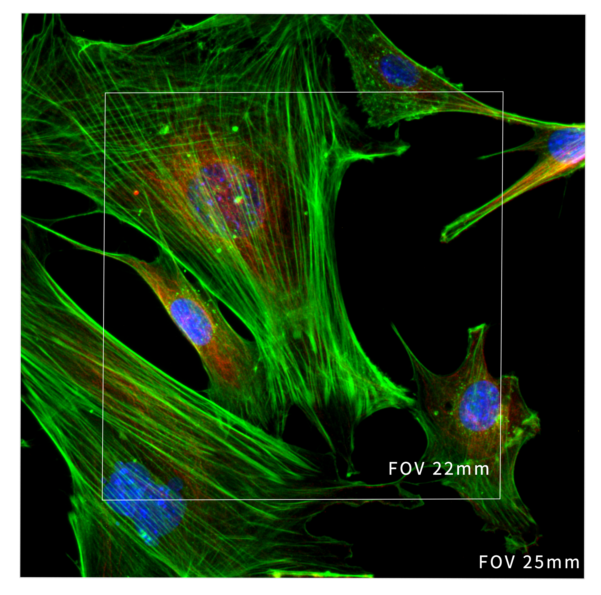
25mm Field of View
The NCF2000 expands the field of view to 25mm, allowing large-format cameras to capture more data with every image.
Key Features

Resonant Scanning - Capture Life in Motion
High-speed imaging up to 60 FPS at 8×256 pixels and 25 FPS at 512×512 pixels significantly reduces exposure time and phototoxicity. Rapid, high-frequency data acquisition enables accurate, real-time capture of dynamic events and prolonged cellular changes.


High-Sensitivity GaAsP Detection
The GaAsP photomultiplier tube improves fluorescence collection efficiency, enabling detection of faint fluorescent samples that conventional methods struggle to capture. Supports four spectral channels across 400-750nm with adjustable precision of 1nm.

NomisProX-C Software
Integrated control of the confocal system and microscope hardware in one platform. Supports multi-channel fluorescence imaging, time-lapse scanning, multi-position acquisition, Z-axis stacking, and panoramic stitching. Built-in 2D and 3D deconvolution further enhances image clarity and resolution.

High-Speed Electric Motor Control
Motorised control of objectives, filter blocks, XY stage, and observation modules creates an effortless operating environment. A joystick provides intuitive stage manipulation, keeping the focus on the experiment rather than the instrument.
25mm Field of View
The NCF2000's industry-leading 25mm FOV captures large-area samples in a single scan, delivering 1.5x the data throughput of the previous generation. Paired with 8192×8192 pixel scanning resolution, it ensures acquisition of vital biological data with exceptional detail and efficiency.


Real-Time Four-Channel Synchronised Imaging
Simultaneously detect and analyse four distinct fluorescent labels within the same field of view. Precise spectral unmixing imaging reveals intricate, multi-layered information within samples, vastly enhancing experimental throughput and data accuracy.

Spatial Array Confocal Scanning Head
An integrated high-precision galvanometer scanning system with a continuously variable electric pinhole delivers low-noise, high-contrast confocal imaging. The 5×5 array single-pixel photon counter achieves ultra-high resolution - 120nm in XY and 350nm in Z.

Super-Resolution Ready
Compatible with the NSR Series super-resolution module, enabling 2D-SIM lateral resolution down to 86nm and 3D-SIM axial resolution of 270nm - extending capability well beyond standard confocal imaging.

Key Specifications
| NCF2000 Laser Confocal Microscope Specifications | |
|---|---|
| Laser | 405nm, 488nm, 561nm, 640nm |
| Standard detector | Wavelength: 400-750nm, 4-channel PMT detection |
| Spectral detector | Wavelength: 400-750nm, detected by a 4-channel PMT (compatible with a 4-channel GaAsP PMT at most), with a spectral range of 400-750nm and an adjustable precision of 1nm |
| Array detector | 5x5 array single-pixel photon counter; achieves ultra-high resolution imaging with image resolution of 120nm in the XY directions and 350nm in the Z direction |
| Transmittance detector | Single channel PMT detector |
| Scan head | Maximum pixel size: 8192x8192; galvanometer and resonant scanner; maximum scanning speed 10 FPS (512x512, galvanometer scanning) and 25 FPS (512x512, resonant scanning) |
| Scan mode | Supports combined use of X, Y, Z, A, and T scanning functions |
| Pinhole | Electric stepless adjustment |
| Confocal field of view | Square inscribed in a 25mm diameter circle |
| Image bit depth | 16 bits |
| Compatible microscope | NIB1000-AT electrically operated research-grade inverted microscope |
| Optical system | NIS60 infinite optical system |
| Eyepiece | 10x, diopter adjustable -5 to +5 |
| Observation tube | 10-40° inclined observation tube, interpupillary distance adjustable 47-78mm |
| NIS60 objective lens | 10x Plan-Apochromat, NA=0.45, WD=4mm |
| 20x Plan-Apochromat, NA=0.75, WD=1.1mm | |
| 40x Plan-Apochromat, NA=0.95, WD=0.19-0.21mm | |
| 60x Plan-Apochromat, NA=1.42, WD=0.25mm | |
| 100x Plan-Apochromat, NA=1.45, WD=0.13mm (optional) | |
| 100x Plan-Apochromat, NA=1.49, WD=0.09-0.16mm (optional) | |
| Nosepiece | Electric six-position nosepiece (with DIC expansion slot) |
| Electric six-position nosepiece with AFS module (optional) | |
| Condenser | Seven-position electric revolving condenser |
| Illumination system | Transmitted: Koehler illumination with LED lighting; Epi-illumination: widefield 4-band LED lighting; 8-position electric fluorescence nosepiece (B, G, U standard); motorised fluorescence shutter |
| Ports | Beam splitter ratios: Eye 100%, Left Port 100%, Right Port 100%, Eye 20% / Right Port 80% |
| Stage | Electric control (grating type); travel range 130x100mm (stage size 445x300mm); maximum speed 25mm/s; positioning accuracy 0.1μm; repeatability 0.5μm; universal stage plate compatible with 35-65mm culture dishes and slides; optional plate holder adapter |
| Focusing system | Electric Z-axis; travel distance 10mm; minimum step 0.02μm; repeatability 0.1μm; three-speed focusing knob (2μm/revolution, 40μm/revolution, 200μm/revolution) |
| DIC slider | 10x, 20x, 40x, 60x, 100x objective inserts, compatible with the nosepiece turret (optional) |
| Software | Nomis Pro X-C; confocal scanning, multidimensional imaging, and pre-set channel functions; comprehensive hardware control modules; built-in image filtering tools; supports export to various image formats |
| Camera | Nexcam-T12 |
| Expandable functions | Live cell culturing system; super-resolution structured illumination microscopy (SR-SIM) module; dual-layer fluorescence heightened accessory |