
New probe from Spirochrome: CenSpark650, the first centriole and cilia selective probe
The first Fluorescent probe for Centrioles and Cilia!
CenSpark650™ is a unique fluorescent live cell probe for centrioles and cilia fluorescence imaging. CenSpark650™ recognizes microtubule triplets and doublets structures present in centrioles and cilia. CenSpark650™ is a bright, far red, fluorogenic, non toxic, cell permeable and highly specific live cell probe for fluorescence imaging of centrioles and cilia. It does not require any transfection and works just by adding the probe to the cells and perform imaging a couple of hours later.
CenSpark650™ is based on our bright, photostable & far red SiR fluorophore which is far superior than fluorescent proteins.
CenSpark650™ enables multicolor imaging with SPY505, SPY555, SPY595, SPY700, GFP or m-cherry. CenSpark650™ can be imaged with standard Cy5 filterset. It can be used for widefield, confocal, SIM or STED imaging of living cells and tissue.
Product contains 1 vial of CenSpark650™ in lyophilised form allowing to prepare 50 ul of 1000x DMSO stock solution (100 stainings*).
*Based on the following conditions: 0.5 ml staining solution / staining experiment with 1x probe concentration. The number of staining experiments can be further increased by reducing the staining solution volume or the probe concentration.
The first Fluorescent probe for Centrioles and Cilia! Now available from Klein Scientific.
We are delighted to announce Spirochrome’s new CenSpark650, a dual-ligand fluorescent probe for centrioles and cilia selective imaging.
Visualising centrioles and cilia in live cells was only available through complex genetic engineering. To overcome this limitation, Spirochrome have developed CenSpark650, a transformative small-molecule probe designed for the selective visualisation of centriolar and axonemal microtubules.
CenSpark650 exploits a unique structural feature: the juxtaposition of inner and outer microtubule-binding sites found exclusively in the microtubule triplets and doublets of centrioles and cilia. CenSpark650 achieves unprecedented selectivity in both live and fixed specimens across diverse eukaryotic systems.
This novel tool enables researchers to:
– Track centriole dynamics in live cells over extended periods with negligible toxicity.
– Visualize primary and motile cilia without the need for genetic manipulation, making it ideal for difficult-to-transfect cells and non-model organisms.
– Monitor rapid cellular events, such as centrosome polarization at the immunological synapse in CAR-T cells, with superior spatial resolution.
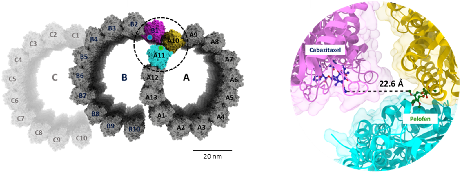
Figure 1. Left: Centriole and cilia microtubule doublets and triplets feature a unique architecture where the “outside” of the A microtubule is in close proximity of the B microtubule lumen. Right: This unique configuration brings in close proximity the binding taxol and peloruside binding sites.
Its highly straightforward “add and image” protocol allows for centrioles to be stained as fast as within 1h. Censpark650™ operates on a unique dual-ligand principle designed to exploit the specific structural architecture of these organelles: it physically couples two different microtubule-binding agents (cabazitaxel and a peloruside A analog) via a long linker, allowing the probe to simultaneously bind to both the inner and outer microtubule-binding sites that exist in close juxtaposition exclusively within the microtubule triplets and doublets of centriolar and axonemal structures (Figure 1)1). This bivalent binding mechanism confers CenSpark650™ with an exceptional selectivity for centrioles over standard, indiscriminate cellular tubulin networks, and because it is fluorogenic, its far-red (670 nm) fluorescence intensity increases only upon target binding, resulting in a highly specific, low-background signal ideal for live cell imaging.
Want to know more? Read the full study published in Nature Chemical Biology: Pourroy, C., Hatzopoulos, G.N., Reymond, L. et al. Development of the fluorescent probe CenSpark for labeling centrioles and cilia. Nat Chem Biol (2026).
CenSpark650™ Probe Properties
| Absorbance maximum λabs | 652 nm |
| Fluorescence maximum λfl | 674 nm |
| Works on fixed cells? | Yes, GA or acetone fixation |
| Probe quantity | 100 stainings* |
| Fluorescence lifetime | 3.0 ns |
| STED depletion wavelength | 775 nm |
| Shipping | room temperature |
| Storage | -20°C |
*Based on the following conditions: 0.5 ml staining solution / staining experiment with 1x probe concentration. The number of staining experiments can be further increased by reducing the staining solution volume or the probe concentration.
Need more information? Contact sales@kleinscientific.com.au